
After a sacral fracture, rehabilitation does not follow a unique pattern. The type of fracture (vertical, transverse, or H-shaped), the patient’s age, and the presence or absence of nerve damage determine both the timeline for weight-bearing resumption and the choice of exercises. Measuring these variables allows for the protocol to be adapted instead of following a generic program.
Sacral Fracture: Resumption Timelines Based on Injury Type
Sacral fractures do not form a homogeneous group. Their location on the bone and the mechanism of occurrence profoundly modify the rehabilitation strategy, particularly the moment when weight-bearing becomes possible again.
| Type of Fracture | Common Mechanism | Weight-Bearing Resumption | Neurological Risk |
|---|---|---|---|
| Vertical (lateral zone) | High-energy trauma (fall, accident) | Relatively early under control | Low if non-displaced |
| Transverse (lower zone) | Sitting fall, direct trauma | Progressive, depending on pain | Moderate (S3-S5 roots) |
| H-shaped (multiple lines) | Osteoporosis, bone insufficiency in seniors | Delayed, strict supervision | High (frequent involvement of sacral roots) |
So-called “H-shaped” fractures are regularly underdiagnosed. According to the ASAF association, they require more advanced imaging (CT scan, sometimes MRI) before planning rehabilitation, as too early a resumption of weight on the pelvis worsens displacement.
A program found online that suggests resuming walking as early as the second week may be suitable for a non-displaced vertical fracture but could prove dangerous for an unstable H-shaped fracture. Imaging diagnosis precedes any rehabilitation decision.
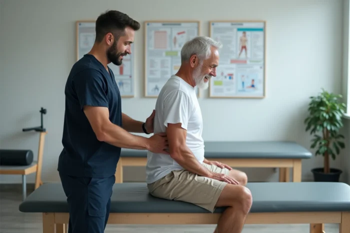
To deepen these distinctions, several resources detail advice for sacral rehabilitation based on the fracture profile and the patient’s background.
Activation of the Gluteus Maximus and Pelvic Stability After Fracture
Most online published sacroiliac rehabilitation programs focus on stretching. Stretching the piriformis or the pigeon pose appears in almost all competing articles. These exercises relieve pain in the short term but do not address the stability deficit that follows a fracture.
The gluteus maximus plays a central role in stabilizing the sacrum. After immobilization, even partial, this muscle quickly loses strength and neuromuscular recruitment. Research referenced by Physiotutors shows that a gradual and staged activation of the gluteus maximus reduces lower back pain associated with sacroiliac dysfunctions.
Staged Progression for Pelvic Strengthening
The logic of progression is based on a simple principle: do not ask the pelvis to stabilize a load before the peri-articular muscles are capable of controlling it.
- Stage 1: floor exercises without load, such as the bridge (two feet on the ground, controlled lift of the pelvis). The transverse abdominal muscle is engaged during exhalation to lock the lumbosacral region.
- Stage 2: unilateral bridge and four-point exercise (opposite arm and leg raised simultaneously), which engages the gluteus medius and deep stabilizers of the pelvis.
- Stage 3: standing exercises with support (lowering onto one foot from a small bench, contraction of the gluteus medius held for several seconds), followed by walking on varied terrain.
The transition from one stage to the next depends on the absence of pain in the sacroiliac joint during and after the exercise. Any acute pain in the sacrum during movement requires a return to the previous stage.
Early Resumption of Walking: What Recent Protocols Change
For a long time, the guideline after a sacral fracture consisted of prolonged strict rest, sometimes several weeks of bed rest. This approach is declining. Several French and Swiss trauma centers now recommend a supervised resumption of walking earlier in the consolidation process.
The goal is not to walk normally within the first few days but to maintain a minimal mechanical stimulus on the bone to promote consolidation. However, this early resumption is only feasible if imaging confirms a stable fracture and no neurological deficit is detected.
Warning Signs During Sacral Rehabilitation
Some symptoms during the rehabilitation phase require immediate medical advice:
- Loss of sensation in the perineal area (bowel), which may indicate involvement of the lower sacral roots.
- Pain radiating into one or both legs, suggesting nerve compression at the pelvic level.
- Sudden worsening of sacral pain after exercise or a change in position, suggesting secondary displacement of the fracture.
These signals do not require an adjustment of exercise but a medical reevaluation. Sacral fractures with neurological involvement represent a minority of cases, but their consequences are serious if ignored.

Sitting Posture and Fractured Sacrum: An Underestimated Parameter
Prolonged sitting exerts direct pressure on the sacrum and coccyx. After a fracture, this daily mechanical strain hinders consolidation if not managed.
A seat cushion hollowed out in the center (such as a coccyx cushion) reduces pressure on the fractured area. Limiting sitting periods to less than thirty consecutive minutes during the first weeks of rehabilitation decreases the cumulative load on the sacrum.
Regularly changing posture (sitting, standing, lying down) maintains mobility of the sacroiliac joint without imposing prolonged static strain. This point, rarely detailed in online exercise programs, conditions the quality of consolidation as much as the exercises themselves.
Rehabilitation after a sacral fracture relies less on the number of exercises performed than on their suitability for the diagnosed fracture type and adherence to progression stages. An initial CT scan or MRI, monitoring of neurological signals, and rigorous management of sitting posture form the foundation on which pelvic strengthening exercises yield lasting results.